
Menu
Contact Information
Principal Investigators:
Prof. Bjørn Torger Stokke
Department of Physics
Høgskoleringen 5
N-7491 Trondheim, Norway
Email: bjorn.stokke@ntnu.no
tel. +47 73593434
Assoc. Prof. Marit Sletmoen
Department of Physics
Høgskoleringen 5
N-7491 Trondheim, Norway
Email: marit.sletmoen@ntnu.no
tel. +47 73593463
Biological polymers: equipment
Atomic force microscopy - Digital Instruments Nanoscope IIIa
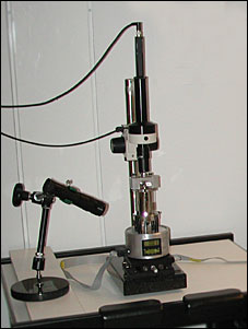
The atomic force microscope is a multimode scanning probe microscope with a NanoScope IIIa controller and a PicoForce force spectroscopy control module from Digital Instruments. It is equipped with three different scanner heads, an "E" scanner with a scan area of 10x10 µm, a "J" scanner that gives the possibility of scanning an area up to 125x125 µm, and a "PF" scanner with a 40 µm xy range and a 20 µm z range. The PicoForce scanner has a new z sensor and is connected to a PicoAngler that gives great advantages in force plotting and pulling of macromolecules. In atomic force microscopy, a microscale cantilever with a sharp tip at its end is used to scan the specimen surface. When the tip is brought into proximity of the sample surface, forces between the tip and the sample lead to deflection of the cantilever. This deflection is measured using a laser and a photo diode detector, and an image of the samples topography is formed.
Atomic force microscopy with TIRF - Digital Instruments BioScope SZ - Zeiss Laser TIRF
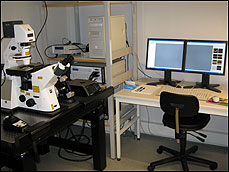
The BioScope SZ is built around the placement of an AFM on an inverted optical microscope, in this case the Axio Observer from Zeiss. This combination simplifies positioning of the sample and finding the area of interest. Applications include pulling or indenting with pico-Newton accuracy, observation of protein folding and unfolding and manipulation of single molecules. The system can be used in both contact and tapping mode, in air and with fluid cells, for which we are equipped with a Fluid Heater that provides temperature control of living cells and other samples. The x-y scan range is 90 x 90 um, and the vertical z range is around 10 um. Included in the system is the NanoMan manipulation module. We have combined the BioScope SZ with a Laser TIRF-system from Zeiss. TIRF (Total Internal Reflection Fluorescence) is used to visualize processes that take place near the cell membrane of living cells, processes that need higher resolution than can be achieved through confocal microscopy. Total reflection occurs at the interface between an optically denser to an optically less dense medium, when a light beam enters at an angle greater than the critical angle. A standing evanescent wave is formed at the transition between these two media, which decays exponentially with the distance from the interface, making the penetration depth not more than 100 – 200 nm. This thin optical section has a high signal-to-background ratio. The laser is a 100 mW multiline argon-ion laser with wavelengths 458 nm, 488 nm and 514 nm.