|
The autonomic nervous system controls involuntary (visceral) functions and has
three divisions. The sympathetic and parasympathetic divisions consist of
two-neuron chains that connect the central nervous system with the smooth
muscles and glands of the viscera, blood vessels, and skin.
The enteric division is a largely independent system that lies in
the walls of the gastrointestinal tract and controls many digestive functions.
The sympathetic system organizes the involuntary responses that anticipate
maximal exertion (in the extreme, the so-called "fight-or-flight" reaction).
Conversely, the parasympathetic system organizes the involuntary responses
that generally reflect visceral function in a state of relaxation.
|
|
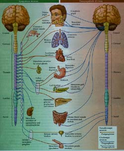
The autonomic nervous system (the enteric division is not shown).
|
|
Sympathetic and parasympathetic ganglia are innervated by preganglionic
neurons in the spinal cord. Sympathetic preganglionic axons arise from neurons
in the thoracic and upper lumbar spinal cord. The preganglionic neurons that
innervate the head and thoracic organs are in the upper and middle thoracic
segments, and those that innervate the abdominal and pelvic organs are in the
longer thoracic and upper lumbar segments. The parasympathetic preganglionic
axons arise from neurons in the brainstem and sacral spinal cord. Many
organs, including the salivary glands, heart, bladder, and sex organs, receive
inputs from both the sympathetic and parasympathetic systems. Other targets
receive only sympathetic innervation. These include the sweat glands, the
adrenal medulla, the piloerector muscles of the skin and most blood vessels. The
neurons innervated by the preganglionic sympathetic axons are for the most part
found in the sympathetic chain ganglia, whereas the parasympathetic motor
neurons are located in ganglia within the organs they control. (The term ganglion
simply means a cluster of nerve cells along the course of a peripheral nerve.)
The enteric nervous system, although it receives sympathetic and
parasympathetc innervation, acts to some degree independently of the rest of
the autonomic system. A rich intrinsic circuitry of sensory neurons, interneurons,
and motor neurons interconnects different levels of the gut and coordinates
activity along its length. Indeed, the enteric system is said to contain more neurons than
the entire spinal cord!
|
|
Abetted by sympathetic and parasympathetic influences, the enteric
system governs gut motility, secretion, and the transfer of substances across the
gut epithelium.
Sensory inputs from the viscera modulate autonomic activity. Like other
primary sensory neurons, the relevant cell bodies lie in dorsal root and cranial
nerve ganglia; the visceral sensory axons that enter the spinal cord terminate
mainly in the intermediate gray matter, near the preganglionic neurons of the
thoracolumnar and sacral cord. Those that enter the brainstem in cranial nerves
VII, IX, and X terminate in the nucleus of the solitary tract, which participates in
many important autonomic reflexes. Sensory fibers that travel in the sympathetic
nerves convey visceral sensations, usually pain. Other fibers, including
most of those that travel in the parasympathetic nerves to the nucleus of the solitary
tract, convey information that does not reach consciousness, but which is
nonetheless important for integration of autonomic reflexes. Examples include
the axons innervating arterial baroreceptors and chemoreceptors. In addition to
mediating the function of the body's glands and visceral muscles, the autonomic
nervous system has provided researchers with a set of relatively accessible
pathways and peripheral preparations that have greatly stimulated neurobiological
research for more than a century.
Teksten er hentet fra boken "Neuroscience", Purves et al. 1997. Sinauer forlag.
E-post til:
Ursula Sonnewald
|